
Fig 3.
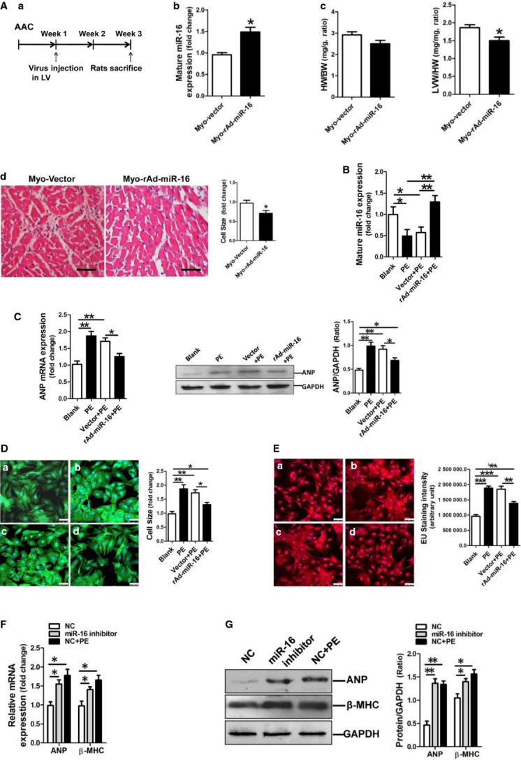
miR-16 inhibits cardiac hypertrophic phenotype in vivo and in vitro. (A) Adenovirus-mediated overexpression of miR-16 inhibited AAC-induced cardiac hypertrophy in rats (n + 6–8). (a) Schematic outline of AAC surgery and study time-points. (b) miR-16 expression in rat myocardium received virus injection was determined by qRT-PCR assay. (c) The ratios of heart weight to bodyweight and LV weight to heart weight. (d) Cell size of cardiomyocytes in rat myocardium received virus injection. miR-16 was overexpressed in the left ventricle, contributing to decreased LV mass. *P < 0.05 versus the vector control. (B) miR-16 expression in NRVCs was determined by qRT-PCR assay (n + 3). (C) ANP mRNA and protein expressions were assessed by assessed by qRT-PCR and Western blotting assay respectively. Restoration of miR-16 resulted in attenuation of PE-induced ANP up-regulation in NRVCs. (D) Cell size assessment of NRVCs by FITC-phalloidin staining assay. (a) Blank, (b) PE treatment, (c) Infection of the adenovirus vector control followed with PE incubation, (d) Infection of the adenovirus expressing miR-16 followed with PE incubation. The scale bar was 50 μm. (E) Assessment of newly transcribed RNA in NRVCs by EU-Apollo staining assay. NRVCs were treated differently in group (a), (b), (c) and (d), consistent with the corresponding groups in the above D. The scale bar was 50 μm. (F) ANP and β-MHC mRNA expressions in NRVCs were determined by qRT-PCR assay (n + 3). miR-16 inhibitor could increase ANP and β-MHC mRNA expressions in NRVCs. NC indicated negative control inhibitor. (G) ANP and β-MHC protein expressions were assessed by Western blotting assay. ANP protein expression was shown increased in miR-16 inhibitor-modified NRVCs. NC indicated the negative control inhibitor. Data are shown as mean ± SD; *P < 0.05, **P < 0.01, ***P < 0.001.