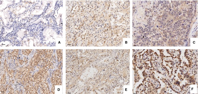
Fig 1.

The expression of LATS1 in LAC examined by IHC staining (×200 and ×400). (A) Low expression of LATS1 in LAC tissues. (B) High expression of LATS1 in ANCT (adjacent non-cancerous tissues). (C) The positive expression of LATS1 was localized in the cytoplasm in LAC. (D) High expression of YAP in LAC tissues. (E) Low expression of YAP in ANCT. (F) YAP was localized in the nucleus in LAC.
