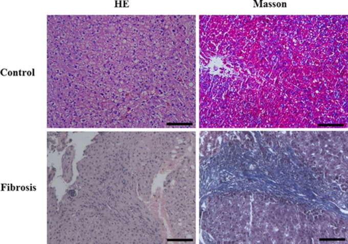
Fig 1.

Haematoxylin–eosin and Masson's trichrome stained sections of human liver. The liver section stained with Masson's trichrome from a control, compared with a liver fibrosis patient showed significant deposition of extracellular matrix proteins. Scale bar + 100 μM.
