Fig 2.
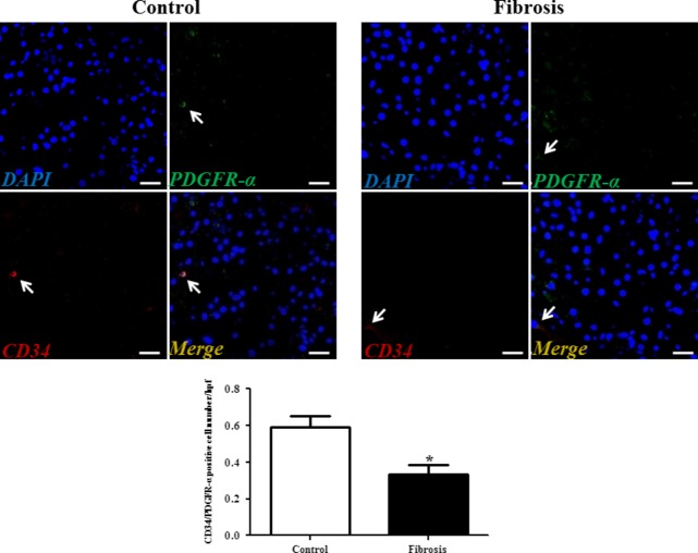
PDGFR-α/CD34 double-positive telocytes are decreased in liver fibrosis. Double immunofluorescence labelling for CD34 (red) and PDGFR-α (green) with DAPI (blue) counterstain for nuclei. Telocytes (TCs) are CD34 and PDGFR-α positive. Scale bar + 50 μm. Quantitative analysis of PDGFR-α/CD34 double-positive TCs in sections of liver from controls and liver fibrosis patients. Data are represented as mean ± SD telocyte number per high-power field (hpf). *P < 0.05, compared to controls.

