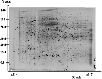
Figure 2.

Representative silver-stained 2DE gel for HepG2 cells without GTT treatment at pH 4–7. The circled and numbered proteins (1a, 2a and 3a) are the differentially expressed proteins. All these three proteins were down-regulated during GTT treatment and could not be detected in the protein profile of HepG2 cells with GTT treatment.
