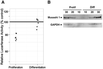
Figure 2.

De-repression of a Musashi-dependent reporter mRNA during differentiation of SH-SY5Y cells. (A) SH-SY5Y cells were transfected with plasmids encoding luciferase reporter mRNAs under control of a Musashi binding element (MBE) or a control 3′ UTR (Control) and luciferase activity was assessed, relative to a co-transfected Renilla luciferase standard, following culture under proliferation or differentiation conditions for 24 hours. The dot plot shows the relative luciferase activity of six independent experiments as a percent of the 3′ UTR control (100%) for both proliferation and differentiation, as indicated. Luciferase expression was repressed in proliferating cells (mean of 77% with a 95% confidence interval: 73 to 81%; p < 0.0001, one sample t-test) but not in differentiated cells (mean of 97.7% with a 85% confidence interval 90.6 to 105.4%, p > 0.47, one sample t-test). (B) Western blot demonstrating persistence of Musashi1 in SH-SY5Y cells 24 hours after induction of differentiation. In this experiment, 10, 20 and 30 μg total protein (as indicted) were analyzed for Musashi1 and GAPDH protein levels from proliferating neurosphere culture (Prolif) or cells cultured in neuronal differentiation media for 24 hours (Diff). A representative experiment is shown.
