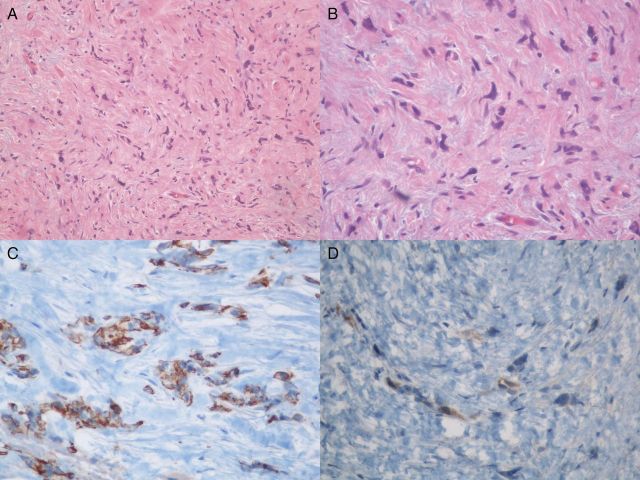
Figure 2:
Histopathological examination results (A) Infiltrating tumoral cells in hyalinized collagenous stroma (H&E × 100) (B) Tumoral cells with spindle, triangular or polygonal shaped and hypercromatic nuclei (H&E × 400) (C) Diffuse positivity with cytokeratin (Clone AE1-AE3 × 200) (D) Scant cytoplasmic positivity with thrombomudulin (Clone 1009 × 200).

