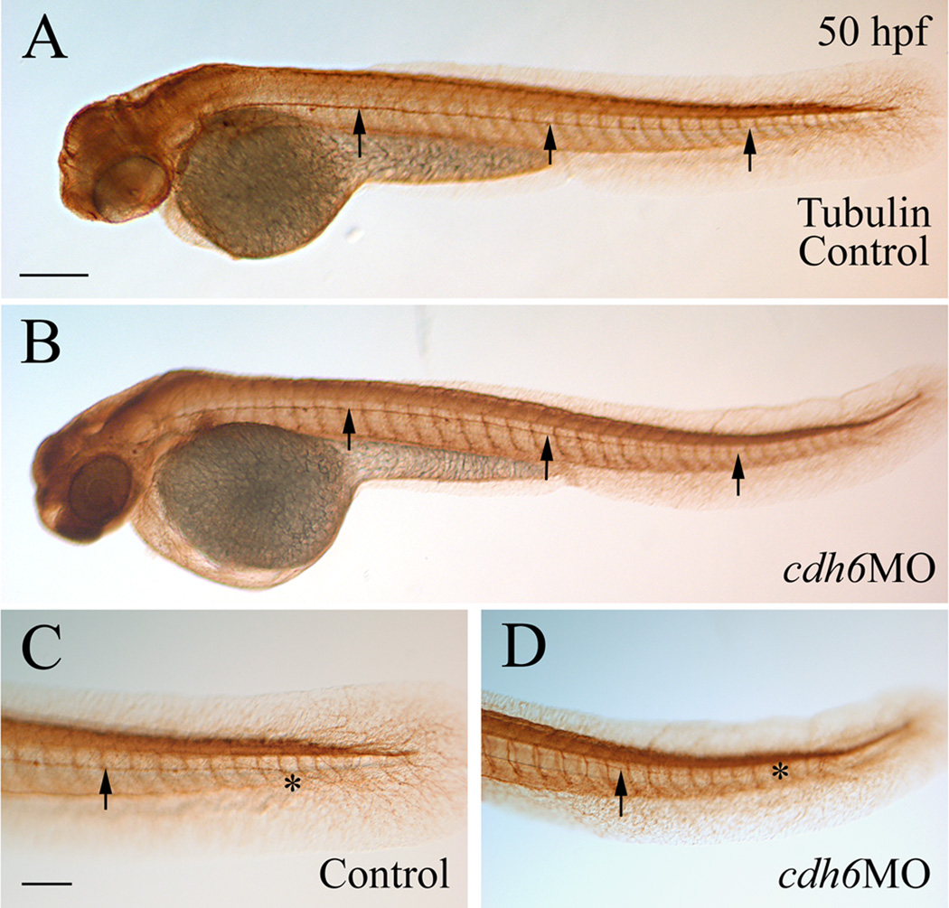
Figure 8.
Normal development of the posterior lateral line nerve in cdh6 morphants. All panels show lateral views of whole mount embryos (anterior to the left and dorsal up) processed for anti-acetylated tubuline immunoperoxidase staining. Arrows point to the posterior lateral line nerve, while the asterisk indicates the terminus of the nerve. Panels C and D are higher magnifications (same magnification) of the tail region of the embryos in panels A and B (same magnification), respectively. Scale bar = 200 µm for panels A and B, 100 µm for panels C and D.

