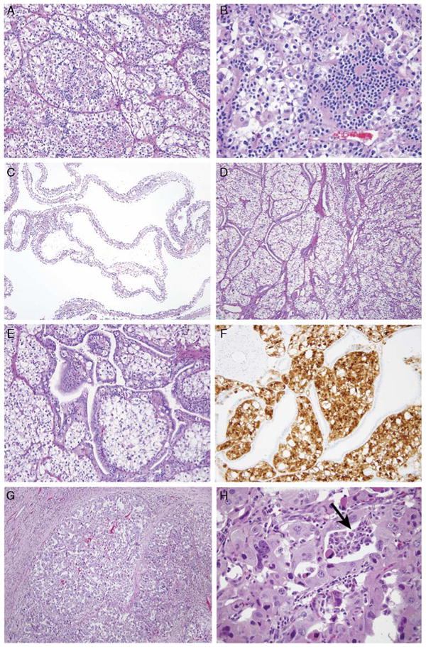
Figure 2.

Typical and unusual morphology of t(6;11) RCC in this study. A and B, The typical morphology of t(6;11) RCC is that of a nested epithelioid cell neoplasm with a second population of smaller epithelioid cells associated with hyaline basement membrane material. C, Case 2 was extensively cystic radiologically and grossly, and microscopically this corresponded to thin cysts lined by clear cells simulating multilocular cystic RCC. D and E, The solid areas of this neoplasm demonstrated nests of clear cells associated with a florid proliferation of entrapped native renal tubules. F, The distinction between the t(6;11) RCC and the entrapped tubules is highlighted on a Melan A stain, which labels the neoplasm but not the entrapped tubules. G, Case 10 was a rather nondescript high-grade nested eosinophilic RCC. H, Very focally, a smaller cell population was present (arrow), which suggested the t(6;11) RCC.
