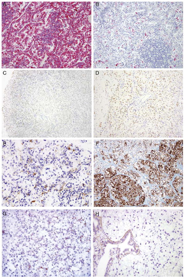
Figure 3.

IHC markers evaluated in the t(6;11) RCC. Melan A labeling was consistently diffuse (A), in contrast to the focal, individual cell labeling consistently seen with HMB45 (B). C, PAX8 immunoreactivity was characteristically stronger at the edge of the sections than in the center, likely representing a fixation artifact. D, At the edge of the tumor distinctive PAX8 nuclear labeling of the neoplasm in the absence of labeling of the intermixed blood vessels was apparent. E, RCC marker labeling was characteristically membranous and distinctly labeled the larger epithelioid cells of a majority of cases. F, The majority of the tumors labeled for CD11 7, including several cases that labeled diffusely. G, A minority of cases labeled for Ksp-cadherin. H, However, the majority of cases were negative, including cases with entrapped native renal tubules, which served as internal controls.
