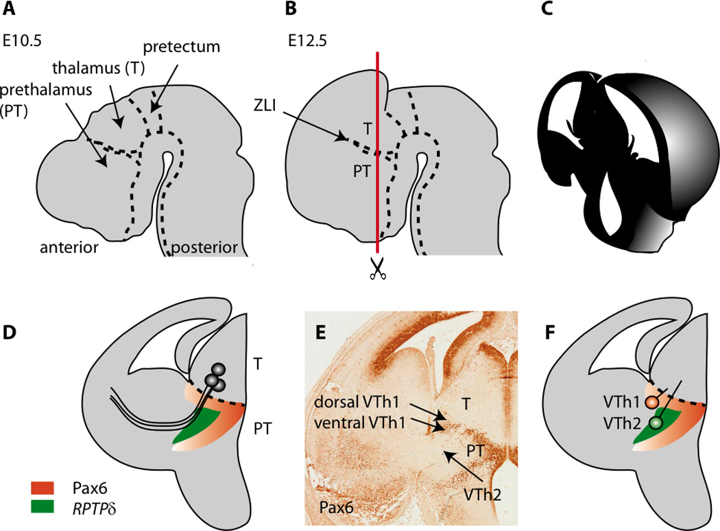
Figure 1.

The anatomy of the developing forebrain and the location of prethalamic cell groups providing guidance for thalamocortical axons in embryonic mouse brain. (A) A sagittal view of the brain around E10.5 showing the pretectal, thalamic and prethalamic anlagen. (B) By E12.5 the telencephalic vesicles expand over the diencephalon; note that the prethalamus (PT) lies anterior to the thalamus (T). These two structures are separated by the zona limitans intrathalamica (ZLI). (C) The appearance of the forebrain when cut as shown by the red line in B at E14 (D) Thalamocortical axons grow from the thalamus, through the prethalamus and into the telencephalon. The prethalamus contains cells that express the markers Pax6 and RPTPδ (Tuttle et al., 1999). (E) An example of a section stained with an antibody for the Pax6 transcription factor (E14). The positions of prethalamic groups of cells proposed by Tuttle et al. (1999) to project to the thalamus and provide guidance to thalamocortical axons are shown. These groups were originally called VTh1 and VTh2, with VTh1 split into a dorsal and a ventral domain. The dorsal domain of VTh1 expresses a low level of Pax6, whereas the ventral domain of VTh1 expresses a high level of Pax6. (F) VTh2 does not express Pax6 but does express RPTPδ.
