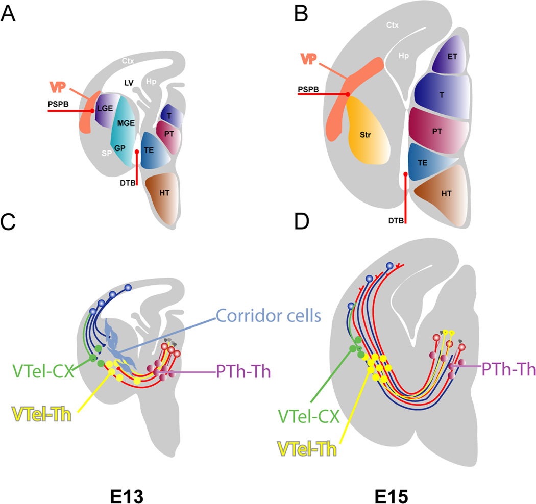
Figure 2.
The position of various cell populations guide thalamic axons. A and B depict the various subdivisions in the diencephalon (ET - epithalamus, T-thalamus, PT-prethalamus, TE-thalamic eminence, HT-hypothalamus) and telencephalon (MGE-medial ganglionic eminence, GP-globus pallidus, LGE-lateral ganglionic eminence, PSPB-pallial subpallial boundary, VP-ventral pallium or SP-subpallium, CTX-cerebral cortex, Str-striatum) and their bounadary (DTB-diencephalic and telencephalic boundary).
C and D issultrates the early connectivity in the telencephalon and diencephalon. Prethalamic (PTh-Th) and ventral telencephalic (internal capsule, VThel-Th) cells with thalamic projections (purple and yellow respectively) are instrumental in the early thalamic axon guidance. Panel in C illustrates the migration of the corridor cells and their interactions with the thalamocortical projections. Corridor cells (light blue) originate from the lateral ganglionic eminence (LGE) at embryonic day 12 (E12) and migrate tangentially toward the diencephalon, where they form a permissive “corridor” for the thalamic projections (red) to navigate them through the internal capsule. Modified based on López-Bendito and Molnár (2003) and Hanashima et al., (2006).

