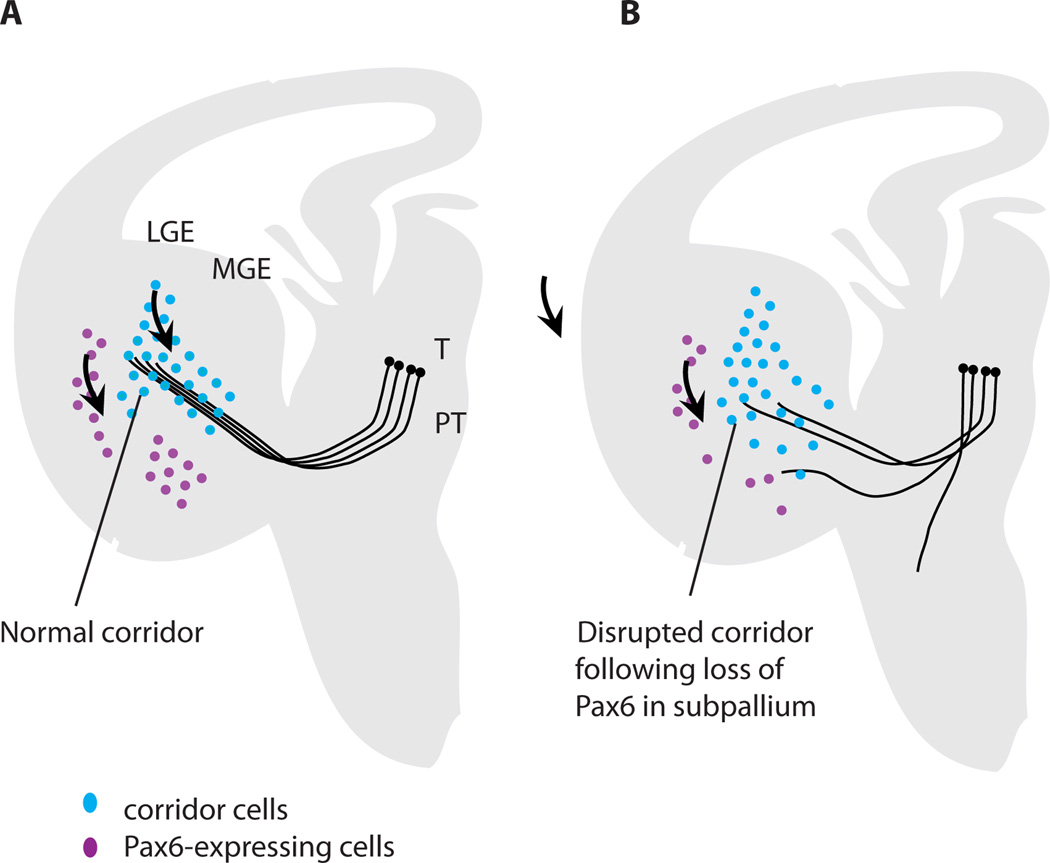
Figure 3.
Evidence that Pax6 plays a role in corridor formation. (A) Normally, Pax6-expressing cells (purple) are located ventral to the corridor/ developing internal capsule in the medial ganglionic eminence (MGE); those located laterally comprise the lateral cortical stream migrating from the pallial-subpallial border (arrow). Islet1-expressing cells (green) migrate from the progenitor layer of the lateral ganglionic eminence (LGE; arrow) to form the corridor through which thalamocortical axons grow (arrow). (B) In conditional mutant embryos with selective reduction of Pax6 in specifically the ventral telencephalon but neither thalamus not cortex, there are fewer Pax6-expressing cells ventral to the corridor than normal (other populations of Pax6-expressing cells outside the region of Pax6 deletion are not shown since they are not affected). Cells from the lateral ganglionic eminence migrate to form a corridor that is abnormally broad with a lower peak density of Islet1-expressing cells; many Islet1-expressing cells stray into the area depleted of Pax6 expression. Many thalamic axons fail to enter this abnormal corridor or exit it along its length. Data are taken from Simpson et al. (2009).

