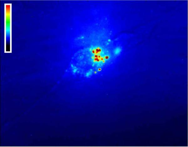
Figure 7.

NIH 3T3 cell in the presence of single-walled carbon nanotubes suspended in E(QL)6EGRGDS. The image shows near-IR fluorescence from the nanotubes, false-colored according to intensity. Bright spots indicate carbon nanotube clusters on the cell surface.
