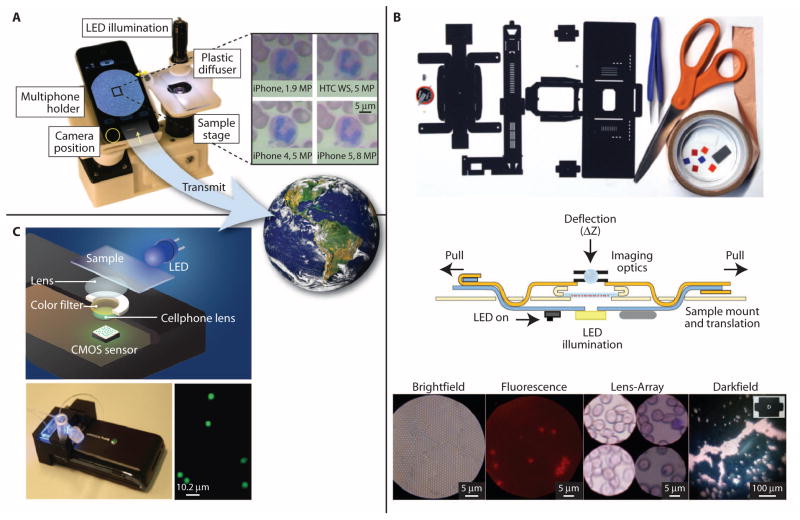
Fig. 2. PoC microscopes and flow cytometers.
(A) A cell phone–based microscope. Four different cell phones were used to obtain images of a Wright-stained blood smear (14), illustrating the resulting differences in image resolution, color, and brightness. For global health care, these images can be viewed locally and transmitted remotely for storage or further analysis. [IMAGE: Visible Earth, NASA.] (B) Foldscope components, tools, and instructions used in its assembly. The cross-section view shows sample mount and translation mechanism, LED illumination, and imaging optics. The sample is inserted from the side. Images were acquired from 1-μm polystyrene beads in bright field, 2-μm fluorescent beads in fluorescence, a Giemsa-stained blood smear in lens array, and 6-μm polystyrene beads in dark field modes (15). (C) Schematic diagram and photo of a cell phone–based flow cytometer. The spatial resolution of the system is about 2 μm, and images acquired with the system show that it can resolve 2- and 4-μm-diameter fluorescent beads (18). Images reproduced from (14, 15, 18) with permission.

