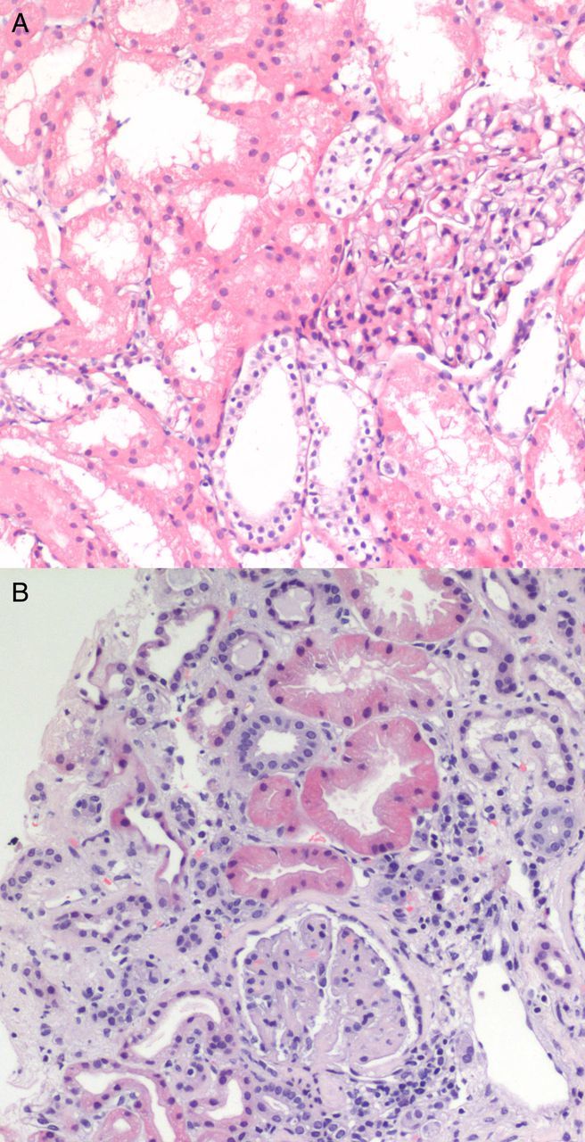
Fig. 1.

(A) First kidney biopsy. Focal proliferative glomerulonephritis, with mesangial matrix and cellular proliferation, and focal hyaline material deposits. No evidence of TMA or endothelial damage. (B) Second biopsy. Advanced TMA, with avascular glomeruli and mesangial sclerosis.
