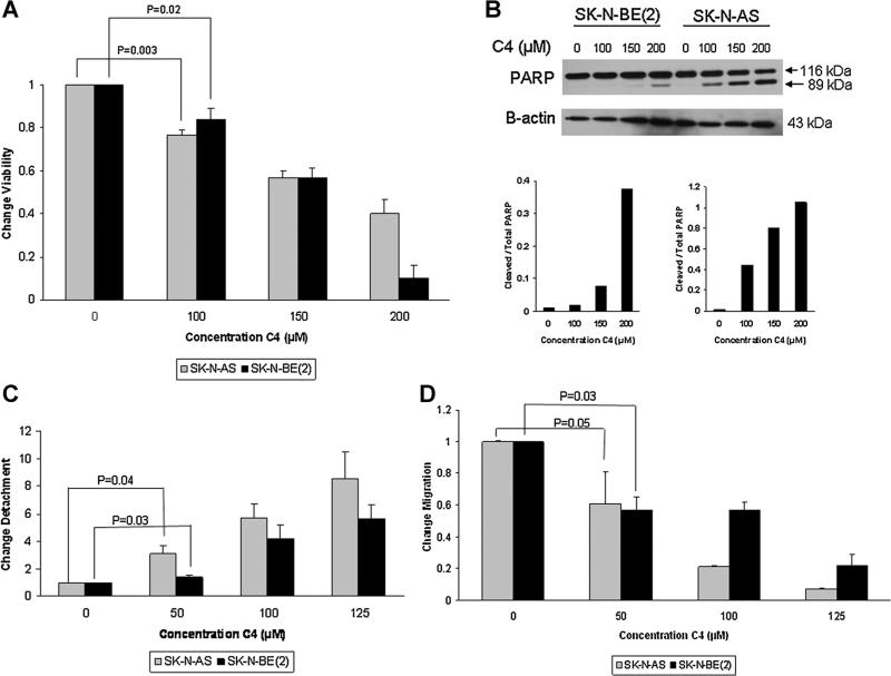
Figure 2.
C4 Decreased neuroblastoma cell viability and led to apoptosis, detachment and decreased migration. (A) SK-N-AS and SKN-BE(2) cell lines were treated with increasing concentrations of C4 for 24 h and cell viability was evaluated with alamarBlue1®® assay. Both cell lines demonstrated a significant decrease in viability after C4 treatment at 100 μM concentration. (B) Both cell lines were treated for 24 h with increasing concentrations of C4. Proteins were separated on SDS–PAGE gels and Western blotting was performed to detect cleavage of PARP, indicating apoptosis. Densitometry was used to further show the differences in cleaved PARP in both the SK-N-BE(2) (lower left graph) and SK-N-AS (lower right graph) cell lines. For densitometry, bands were normalized to β-actin and cleaved PARP compared to total PARP, further demonstrating the increased cleaved PARP following C4 treatment and indicating that the decreased survival was secondary to apoptosis. (C) SK-N-AS and SK-N-BE(2) cells were treated with C4 at increasing concentrations for 24 h. Cell detachment was determined by counting with a hemacytometer and dividing the number of detached cells by the total number of cells (attached + detached), and presented as fold change in detachment. C4 caused significant cellular detachment in both cell lines at 50 μM concentration. (D) SK-N-AS and SK-N-BE(2) cell lines were treated with increasing concentrations of C4 and allowed to migrate through a micropore insert. Migration was reported as fold change in number of cells migrating through the membrane. Cellular migration was significantly decreased in both cell lines with C4 treatment, and as seen with detachment, the effects of C4 upon migration were apparent at 50 μM concentration. The effects of C4 upon detachment and migration were seen at concentrations well below those that affected survival.

