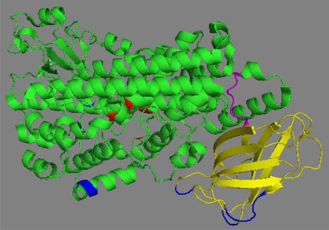
Fig. 4. Crystal structure of the stabilized version of human ALOX5.
The N-terminal β-barrel domain is shown in yellow, the flexible inter-domain linker (D113-L118) in magenta, the C-terminal catalytic domain in green and the iron liganding residues in red. The residues mutated in wild-type ALOX5 to get the stabilized version of the enzyme suitable for crystallization are indicated in blue. The image was constructed from the X-ray diffraction data using the PyMol software package.

