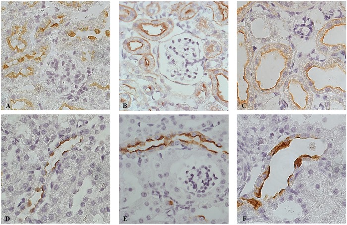
Fig 5. Tubular dilatation mainly involves the distal segments of the nephron.
Representative pictures of renal cortex from Ctr mice (A, D) and Dicer cKO at P30 (B, E) and at P50 (C, F) mice stained with LAH (Lectin from Arachis Hypogaea) (A- C) and AQP2 (D- F). Dilated tubules in P50 Dicer cKO mice are positive for LAH (C) and AQP2 (F), reflecting their origin from distal convolute tubules, connecting and collecting ducts. (A-F) Magnification 63X.

