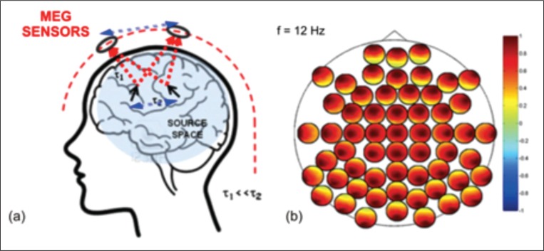
Figure 2.
Schematic representation of signal propagation from brain sources to MEG sensors (a) and of channel-level interactions for a subset of MEG channels (b).
a) signal propagation to the sensors is assumed to be instantaneous in comparison to the timescales of signal propagation within the brain (τ1<<τ2); at a given time instant, the different MEG sensors capture a weighted sum of the activities of all brain sources. b) Schematic representation of channel-level interactions for a subset of MEG channels. The larger black circle indicates the system layout and each smaller circle indicates the coupling of one sensor (black dot) with all the others. The spread of the source activity to the sensors artificially enhances the degree of coupling between channels independently of the actual brain source interaction. Indeed, in this toy example all channels appear to be highly coupled with all the others although only two interacting sources were simulated (a).

