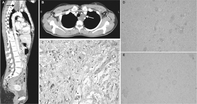
Figure 1.
Radiological, pathological and immunohistochemical features of diagnosed ganglioneuroma.
A and B: Whole-body CT scan showing an expansive lesion in left superoposterior mediastinum (arrows).
C: Hematoxylin-eosin staining of resected ganglioneuroma (original magnification ×40).
D and E: Patient’s ganglioneuroma sections exposed to patient’s serum (diluted 1:20, D) or control serum (diluted 1:20, E). (original magnification ×20).

