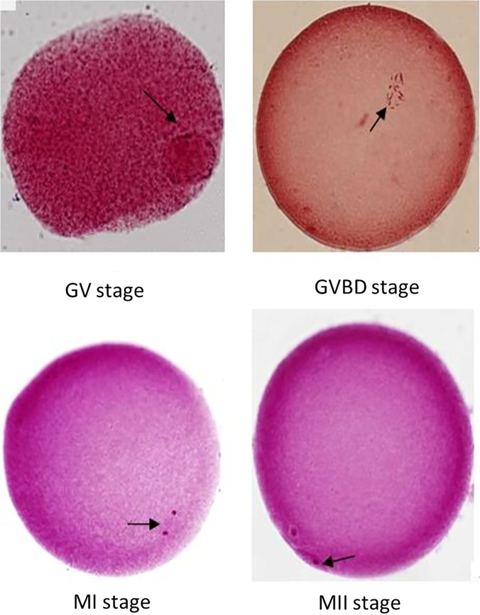
Fig 1. Microscopic characteristics of sheep oocyte maturation stained with aceto-orcein (200× magnification).
(A) Oocyte at GV stage, arrow points to germinal vesicle; (B) oocyte at GVBD stage, arrow points to condensed chromosomes; (C) oocyte at late spindle stage, arrow points to spindle fibers; and (D) oocyte at middle MII stage, arrow points to the first polar body.

