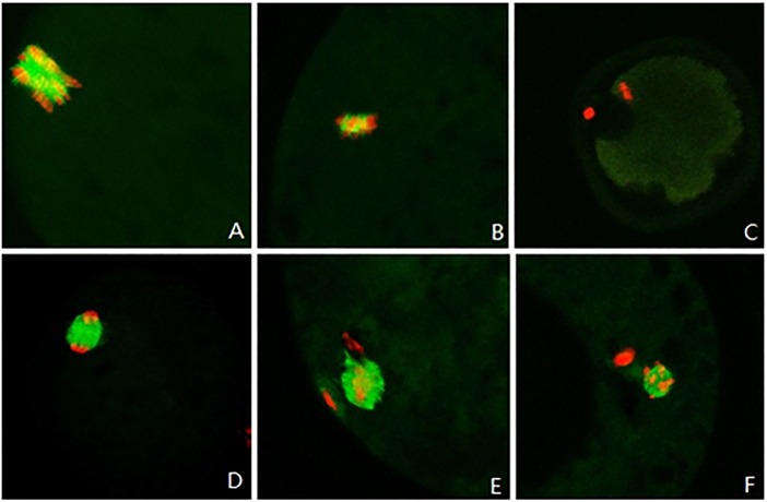
Fig 3. The α-tubulin distribution around chromosomes in sheep oocytes after 22 h of in vitro maturation.
Red indicates chromosomes and green indicates α-tubulin. (A, 10×40) Disorderly distribution of chromosomes; (B, 10×40) little α-tubulin distributed around chromosomes; (C,10×20) nearly no α-tubulin distributed around chromosomes; (D, 10×40) α-tubulin distributed on a spindle; and (E, F, 10×40) formation of microtubules and extrusion of polar bodies at meiosis II.

