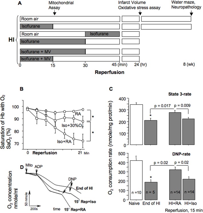
Fig 1. Experimental design.
Mitochondrial phosphorylating respiration rates after HI. (A)—HI-mice, upon reperfusion, were exposed to either room air or isoflurane with or without mechanical ventilation (MV) for different time of reperfusion. (B)—Changes in SaO2 in naive mice (n = 6), and mice exposed to 2 Vol% isoflurane with (n = 6) or without (n = 4) 30% oxygen supplementation, * p < 0.01. (C)—Mitochondrial phosphorylating and uncoupled respiration rates in naïve mice (n = 10), HI-mice at the end of HI-insult (n = 5), and at 15 minutes of reperfusion under isoflurane (HI+Iso, n = 14) or without (HI+RA, n = 14) isoflurane anesthesia. (D)—Representative cerebral mitochondrial respiration tracings from HI-mice tested at the end of HI (End of HI) and 15 minutes of reperfusion with isoflurane exposure (15’ Rep+Iso) or room air (15’ Rep+RA).

