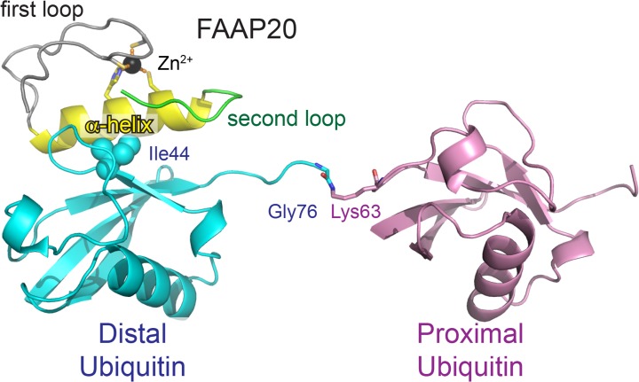
Fig 1. Crystal structure of FAAP20-UBZ in complex with K63-Ub2.
Zinc-coordinating residues (Cys147, Cys150, Cys166 and His169) of FAAP20-UBZ and the isopeptide linkage (Gly76[Ubdistal]–Lys63[Ubproximal]) are shown as sticks. The proximal and distal Ub moieties are colored pink and cyan, respectively. The first and second loops of FAAP20-UBZ are colored gray and green respectively. The central α-helix of FAAP20-UBZ is colored yellow. Ile44 of the distal Ub is shown as spheres. The coordinated zinc ion is shown as a black sphere. Hydrogen bonds are indicated as dashed orange lines.

