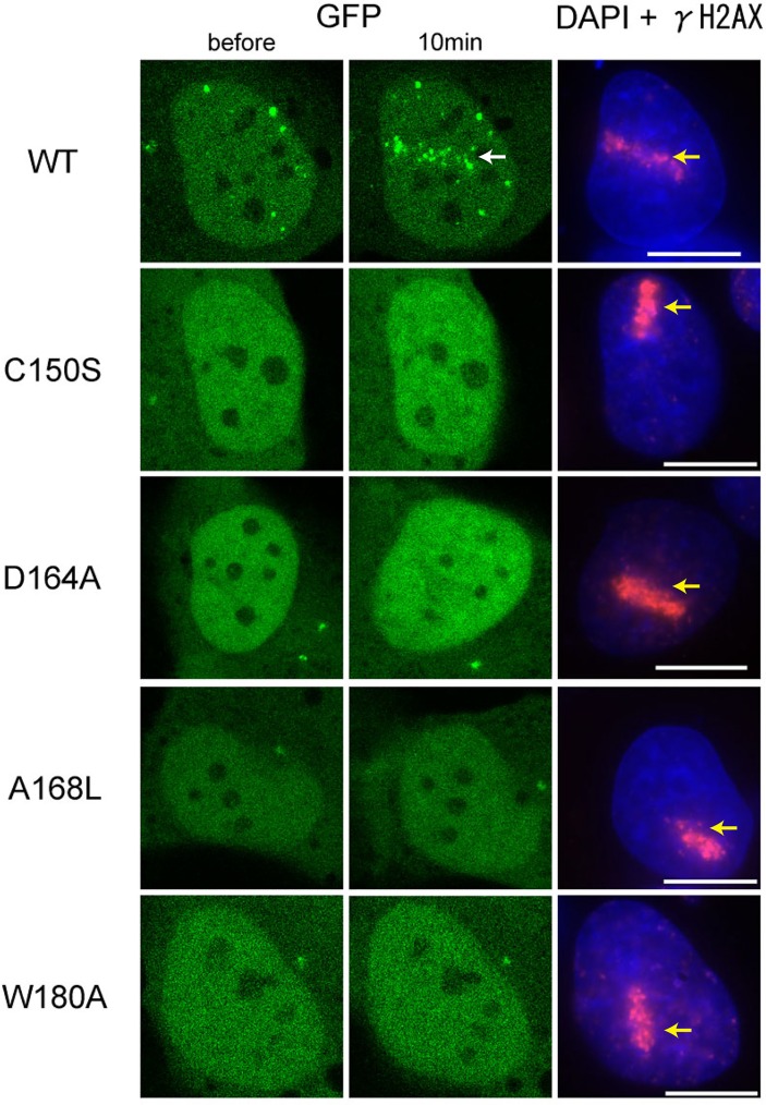
Fig 3. Recruitment of wild-type and mutant FAAP20 to ICLs.
GFP-tagged FAAP20 proteins were detected before and 10 min after laser induced ICLs formation, by confocal imaging. The white and yellow arrows indicate the accumulation of FAAP20 and the γH2AX-positive DNA damage sites, respectively. Scale bars: 10 μm.

