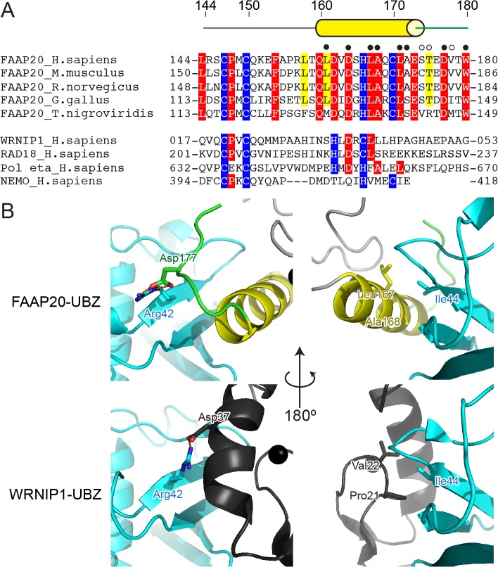
Fig 4. Amino-acid sequence comparison of Polη-, NEMO- and FAAP20-UBZs.
(A) Sequence alignment of five vertebrate FAAP20-UBZs (human, mouse, rat, chicken and puffy fish) and human WRNIP1, RAD18, Polη- and NEMO-UBZs. 100% and more than 80% identical residues among the FAAP20 protein are highlighted by red and yellow background, respectively. Zinc-ion-coordinating residues are highlighted in blue. Open or filled circles indicate the residues that interact with Ub through their main or side chains, respectively (B) Structural comparison of FAAP20- with WRNIP1-UBZ. The coloring scheme is the same as that in Fig. 1. WRNIP1-UBZs are colored dark gray.

