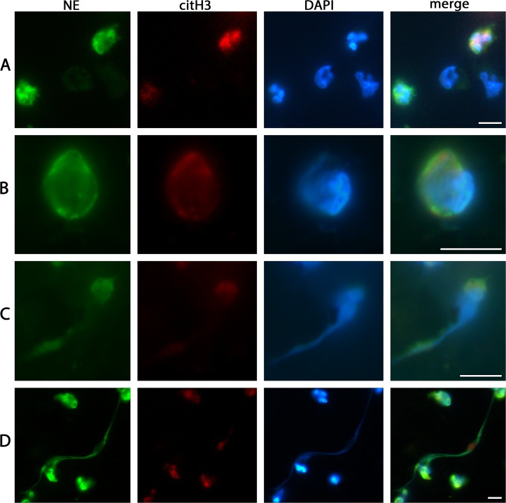
Fig 2. Characteristics of cells adhered to SLA surface from whole peripheral blood as detected by immunofluorescence.
(A) NE-postitive PMNs with their typically lobulated nuclei, and other NE-negative nucleated cells. (B) A single PMN committed to the NETotic cascade as clearly shown by decompensated chromatin, the swollen, partly disrupted nucleus and NE and citH3 staining co-located with chromatin and at cytoplasmic locations. (C) Chromatin extrusion from PMN. (D) Fully spread NETs between PMNs of different activation states. Scale bars: 10μm.

