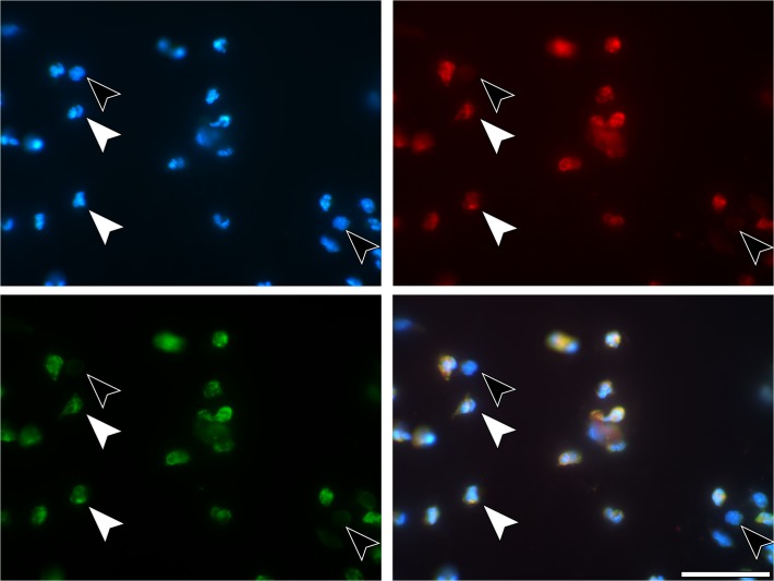
Fig 3. Immunostaining of neutrophils.
(A) 4h SLA sample. PMNs (white arrows) und others, not further determined, cells (black arrows). Blue—DNA staining with DAPI, (B) red—immunostaining for CitH3, (C) immunostaining for neutrophil elastase, (D) merged. The other cells lack both NE and CitH3. Scale bars: 50μm.

