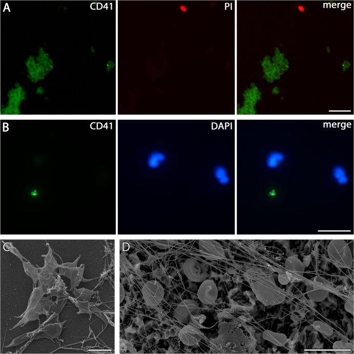
Fig 5. Immunostaining of platelets (A) Detection of platelets adhered from whole peripheral blood to PDL-coated cover slips incubated for 5 min by immunolabelling against CD41 (green).
Large clusters of cells can be found accompanied by few individual nucleated cells (red—PI). Scale bar 10 μm. (B) Platelets staining for CD41 (green) on SLA surface incubated for 5 min with whole peripheral blood are scarcely present between adhered PMNs as shown by lobulated nuclei (blue—DAPI). Scale bar 20 μm. (C) Platelets shown by SEM on uncoated glass cover slips incubated for 4 h with whole peripheral blood. Scale bar 5 μm. (D): SLA incubated for 4 h with whole peripheral blood show no platelets, but numerous erythrocytes as well as fine fibres with fibrin-like and NET-like morphology. Scale bar 10 μm.

