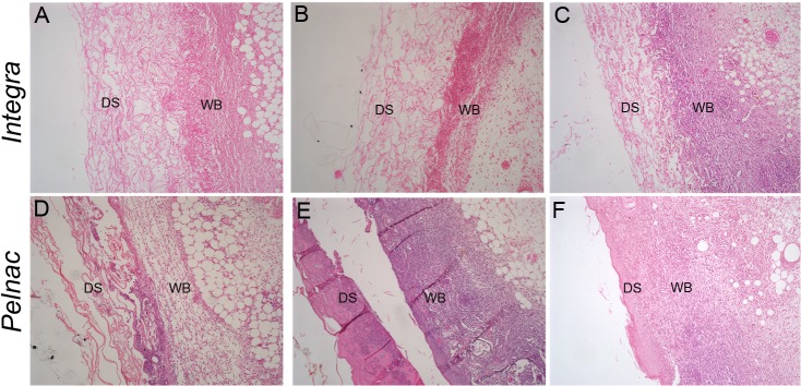
Fig 1. Histological aspects of wounds treated with dermal substitutes.

HE-stained transversal sections of a full-thickness skin wound treated with (A-C) Integra or (D-F) Pelnac collected at (A, D) 3, (B, E) 6 and (C, F) 9 days after the procedure show a clear distinction between the dermal substitute (DM) and the wound bed (WB). Original magnification 100X.
