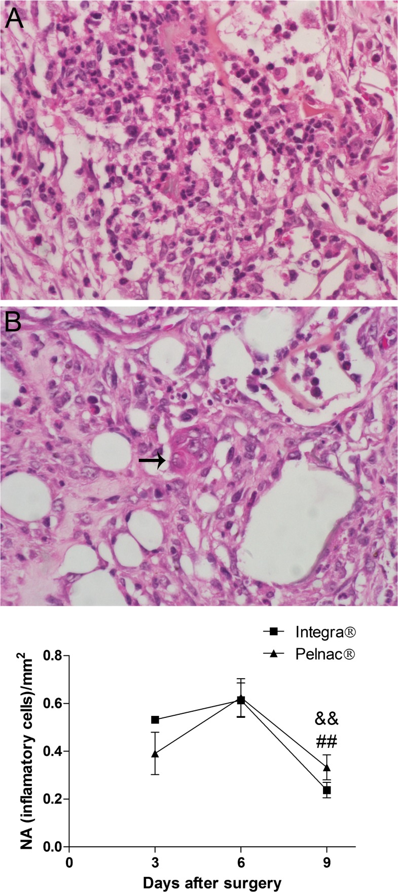
Fig 3. Histological aspects of wounds treated with dermal substitutes showing inflammatory cells.
(A) Representative picture of HE-stained transversal section of a full-thickness skin wound. (B) Foreign-body giant cell (arrow). (C) Quantification of inflammatory cells during the 9 days after the surgical procedure. NA numerical density that represent the number of inflammatory cells per mm2. Data are expressed as the mean ± SEM of 5 animals. Not significant differences between the two templates were observed at any time point. && p = 0.001 Integra at day 6 vs. Integra at day 9 and ## p = 0.004 Pelnac at the day 6 vs. Pelnac at day 9 by two-tailed unpaired t-test. Original magnification 400X.

