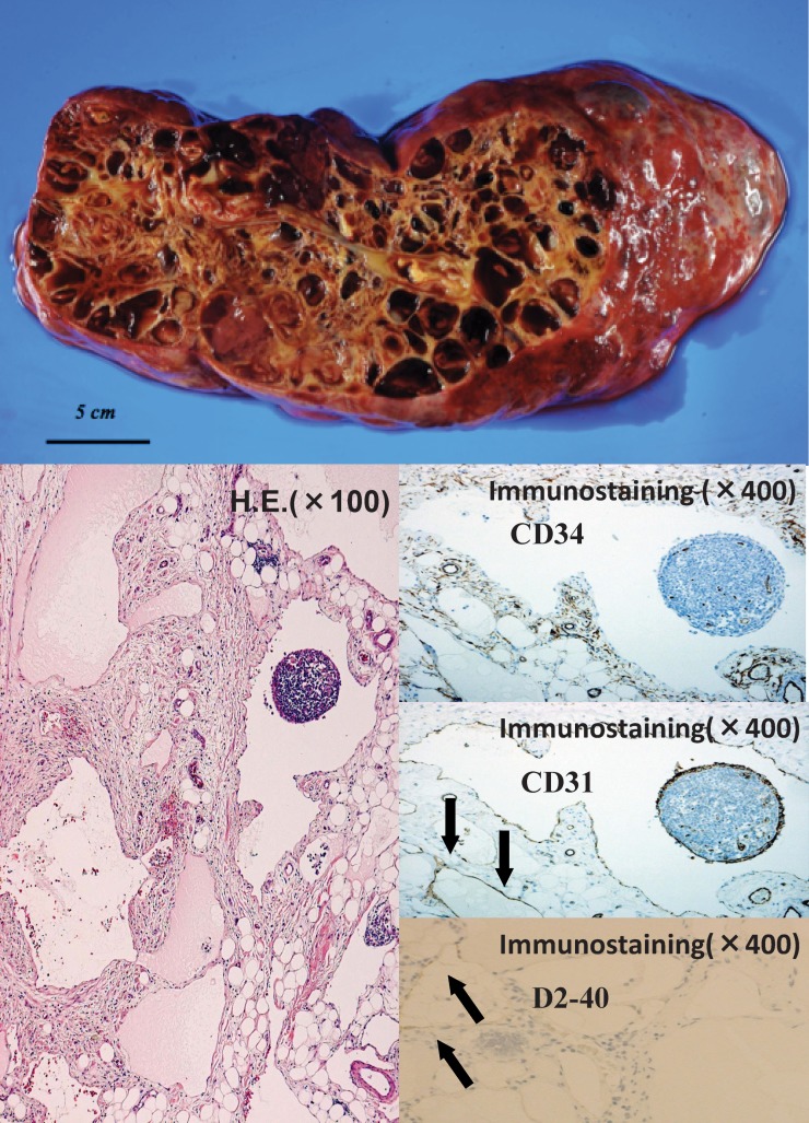
Fig. 5.
Macroscopically, the cystic tumor had many septa and contained yellowish, transparent fluid. A histologic examination revealed variably sized, markedly dilated lymphatic channels in the mesentery, and all parts of the intestinal mucosa were covered with a single, flat layer of endothelial cells. These endothelial cells were positive for CD31 and D2-40, but not for CD34.

