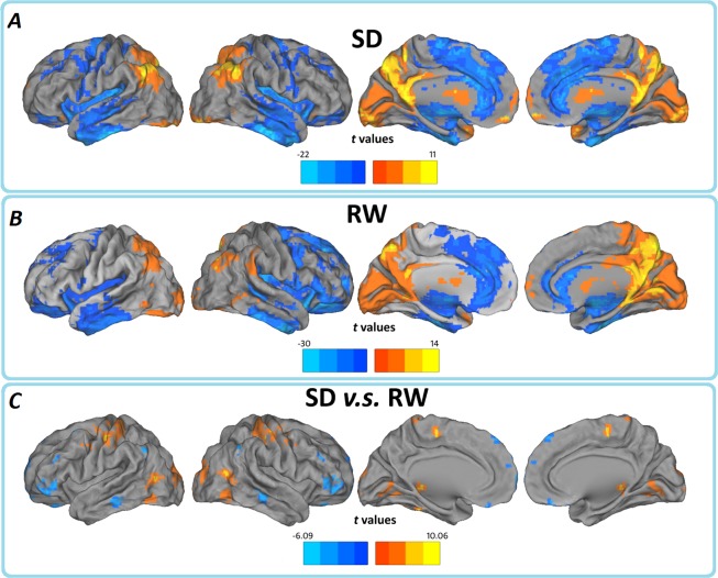
Fig 1. In typical frequency band (0.01–0.08Hz), regions of significant ALFF in the (A) SD and (B) RW groups separately, and their (C) between-group differences.
The effects are significant at p < 0.05, FDR corrected; ≥100 contiguous voxels for one sample t test, and p < 0.05, AlphaSim corrected for paired t test. In the paired t test, cool color indicates that the SD group had decreased ALFF compared with the controls and the hot color indicates the opposite. Left in the figure indicates the left side of the brain.

