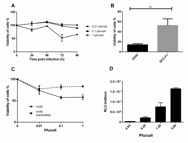
Fig 1. In vitro cytotoxicity and and transduction of vvdd in SCCF1 cells.
A) SCCF1 cells were infected with vvdd with 0.01, 0.1 or 1 pfu/cell. Viability of cells was measured on day 3. B) Compared to A549 cells, SCCF1 cells maintained their viability significantly better on day 3 C) When SCCF1 cells formed a tight monolayer before infection, the cells were still alive 10 days after infection. D) SCCF1 cell were infected with 0.04, 0.2, 1 or 5 pfu per cell and luciferase expression was measured 4 h postinfection in relative light units.

