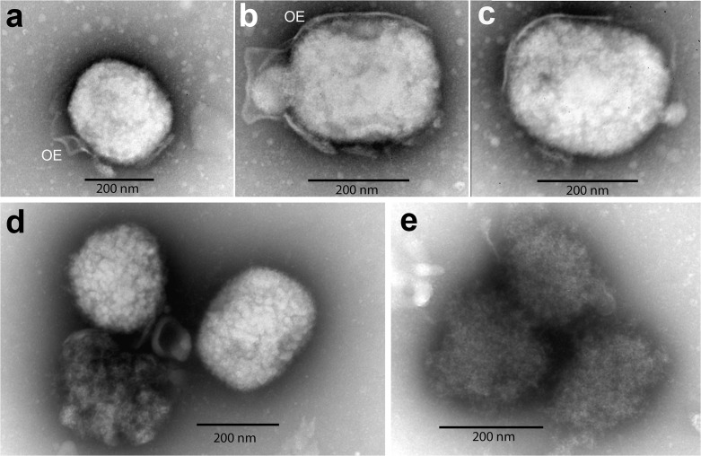
Fig 3. Electron microscopy of negative stained, purified vaccinia viral particles released from cultured cells.
Supernatant from infected SCCF1 or A549 cells was collected, purified and negative staining electron microscopy was performed. A-C: viral particles produced by A549 cells: mature particles of brick-shaped, with outer envelope (OE) derived from cell membrane. D-E: immature vaccinia viral particles produced by SCCF1 cells: the particles are irregular in shape and in electron density. Their internal contents are not tightly packed, no clear boundary or membranous outer envelope is visible.

