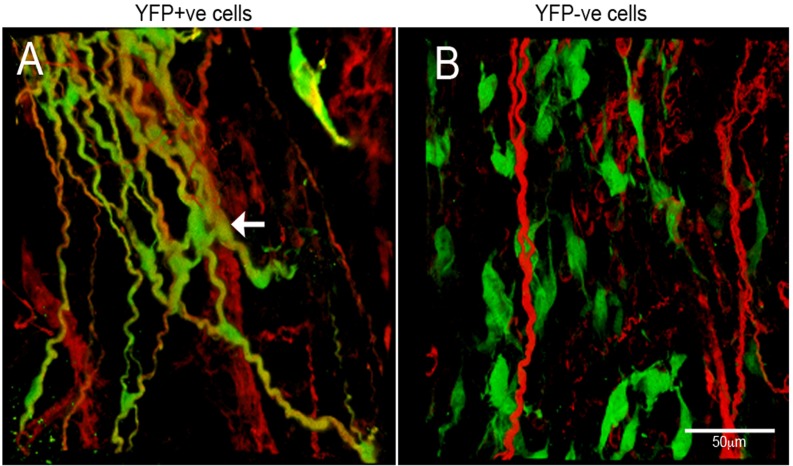
Fig 6. Transplantation of neurospheres into mouse gut.
(A) Confocal images of host gut transplanted with YFP+ve neurospheres. YFP+ve cells spread from the site of transplantation and integrated with the endogenous ENS as shown by TuJ1 immunolabelling (red). (B) Host gut transplanted with YFP-ve neurospheres. YFP-ve cells, labelled with lentiviral construct expressing GFP (green), did not integrate with the endogenous ENS (red) and were apparent as individual, isolated cells. Scale bar = 50μm.

