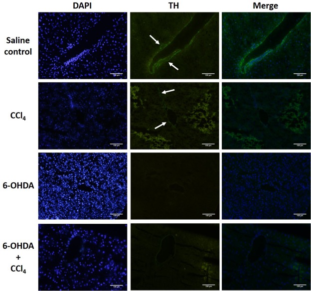
Fig 2. Immunofluorescent analysis of liver tyrosine hydroxylase (TH) reactive nerve fibers.
TH positive nerve fibers (arrows) were seen around the portal area in saline treated mice but not in chemical sympathectomised animals. Magnification × 200. The results are representative of four sets of experiments. Scale bar = 100 μm.

