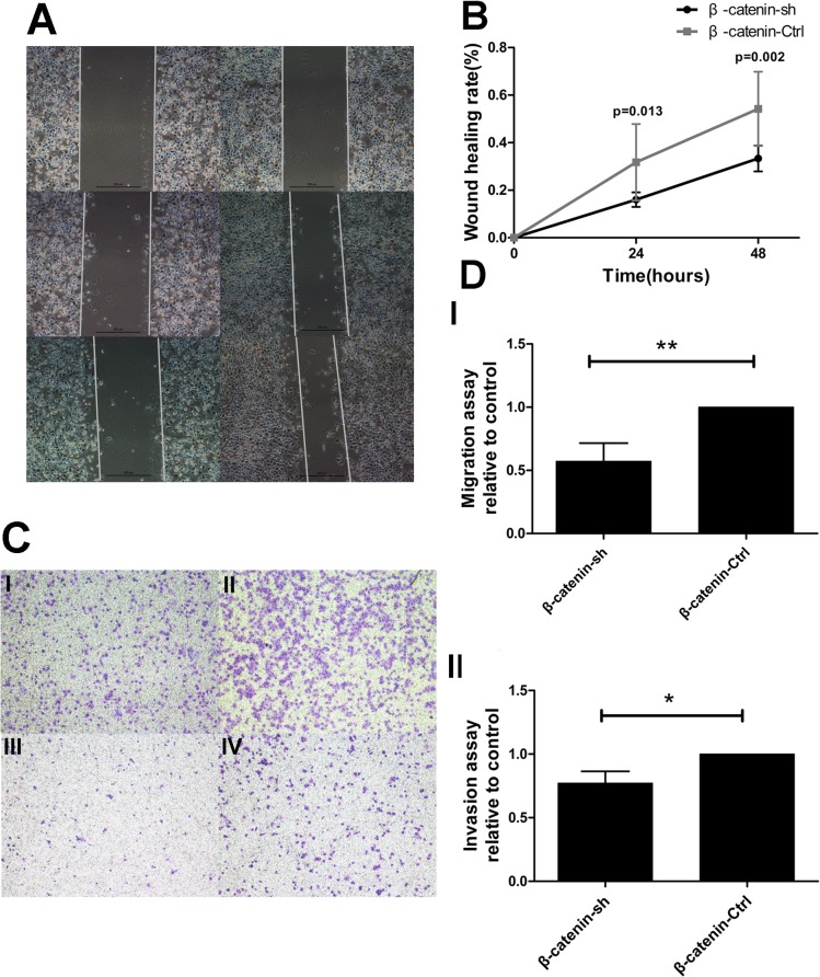
Fig 3. β-catenin shRNA alters the migration and invasion of PDAC cells in vitro.
(A) The wound-healing assay revealed a significant decrease in migration in the β-catenin shRNA group (left), relative to the control group (right). (B) The wound-healing rate was significantly lower in S100A6-expressing cells, compared to the control, at 24 and 48 h. (C) The transwell assay demonstrated that β-catenin shRNA dramatically decreased the number of migrating and invading cells (×40). “I” represents β-catenin shRNA in the migration assay; “II” represents the control group in the migration assay; “III” represents β-catenin shRNA in the invasion assay; “IV” represents the control group in the invasion assay. (D) Representative statistical chart for the transwell assay, showing a significant decrease in migration (I) and invasion (II) in the β-catenin shRNA group, compared to the control group. *p < 0.05; **p < 0.01.

