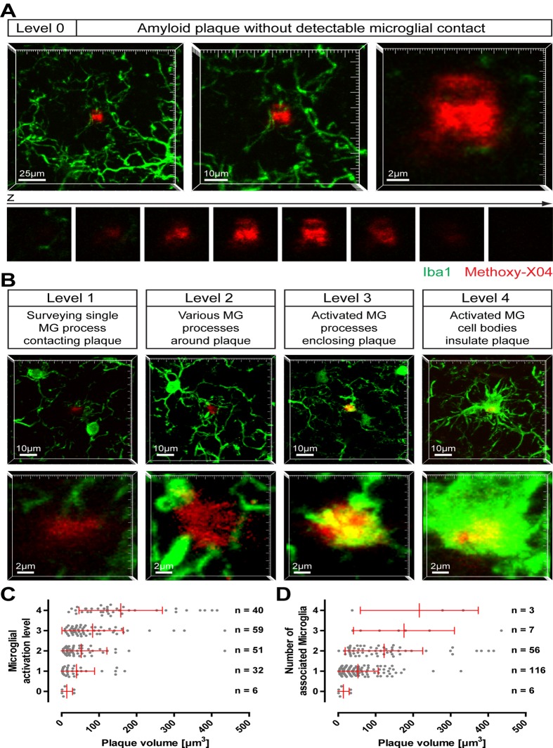
Fig 1. Activation of microglia by small amyloid plaques.
On brain slices of 12-month- old APP-PS1(dE9) mice microglial cells were immunohistochemically labelled for Iba1 (green) and amyloid plaques were stained with Methoxy-X04 (red). Small amyloid plaques were imaged and classified according to the microglial status. A Image projections of an amyloid plaque lacking any microglial contact (level 0) at three magnifications. Below, the single x-y-planes clarifying that no microglial process touches the plaque. B Exemplary images of the stages of plaque-associated microglial (MG) activation (level 1 to level 4). The upper lower magnification illustrates the microglial environment around the plaque, the higher magnification below highlights the individual plaque. Notice that, although each plaque has a similar size, microglial reaction is diverse. C Small plaques were classified according to their microglial activation level and plotted against the plaque volume. D The number of microglial cells associated to the individual plaques was plotted against the plaque volume. (The brains of five animals were analysed with a total of 188 small plaques. Red bars show mean with standard deviation.)

