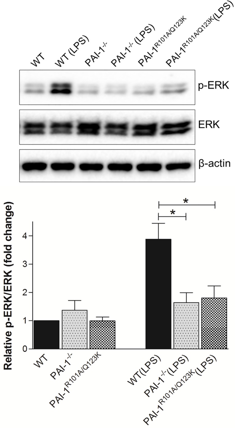
Fig 5. LPS-induced ERK signaling in kidneys 24 hr after LPS challenge.

Protein extracts from renal tissues were prepared as described, and then subjected to Western blot analysis of total ERK1/2 (ERK) and phosphorylated ERK1/2 (p-ERK). β-actin was used as a loading control. The relative p-ERK expression was determined by densitometric analysis (lower panel). Data are expressed as the mean ± SEM (N = 5–6 mice per group), *P<0.05.
