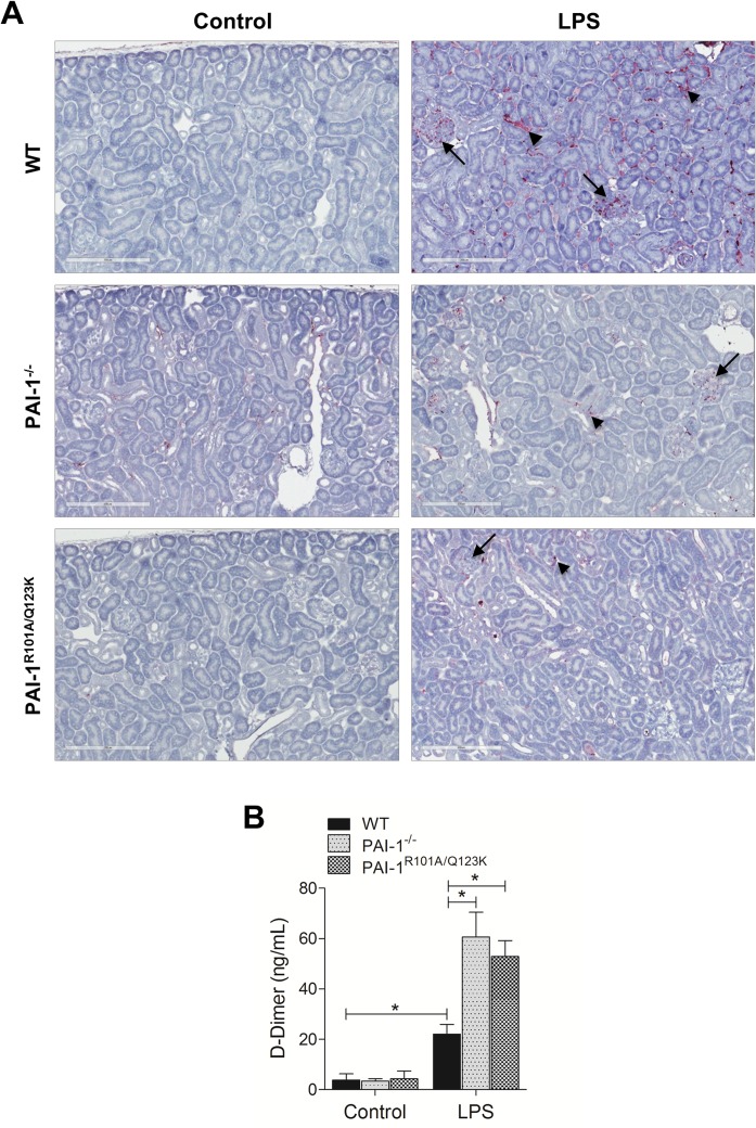
Fig 6. Fibrin deposition in the kidneys 24 hr after LPS challenge in mice.
(A) Fibrin deposition in the kidney. Kidney specimens were subjected to immunohistochemical analysis. Fibrin deposits (purple) in both glomeruli (long arrows) and tubules (small arrows), are indicated. Magnification: 20x. (B) Plasma levels of D-dimer. Data are expressed as the mean ± SEM (N = 4 mice per group), *P<0.05.

