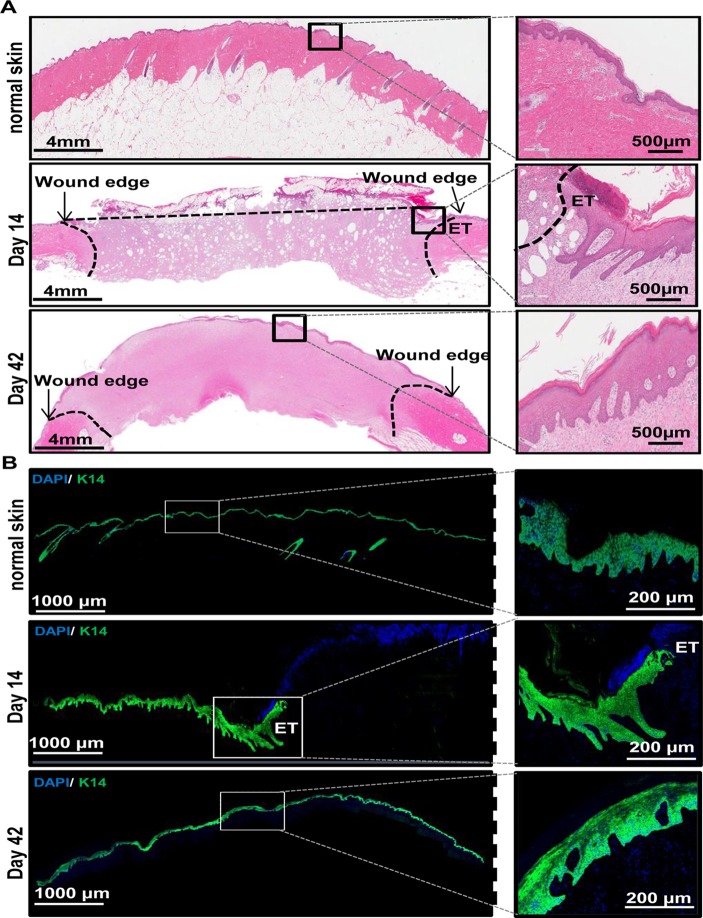
Fig 2. Histological characterization of wound healing.
(A) Shown are representative images (left panels) from formalin-fixed paraffin-embedded biopsy tissue sections (10 μm) of normal and wounded skin (days 14 and 42) that were immunostained using hematoxylin (blue) and eosin (red). Zoomed in images (right panels) of areas in each sections are also shown for better visualization of the epithelial layer of the skin. (Scale bar = 4 mm (left panels) or 500 μm (right panels)). (B) OCT embedded frozen wound biopsies were sectioned (10 μm) and stained using anti—keratin-14 (green) and DAPI (blue). Shown are representative images (left panels) of the stained tissue sections from normal skin and wounded skin (days 14 and 42). Also shown are zoomed in images (right panels) of areas in each section for better visualization of K14 stained epithelial layer of the skin. ET = epithelial tongue. (Scale bar = 1000 μm (left panels) or 200 μm (right panels)).

