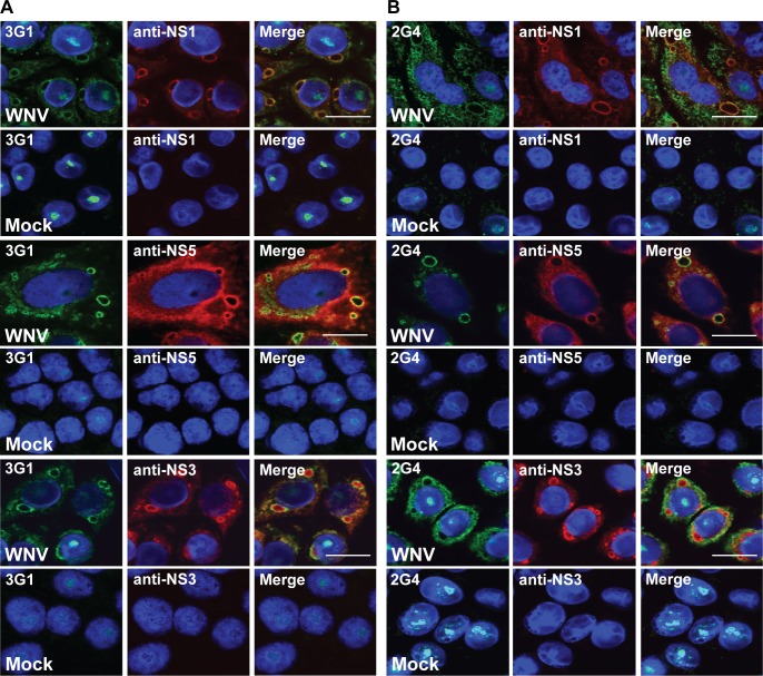
Fig 5. Co-localisation of mAb 3G1 and 2G4 staining with flaviviral replication complexes.
Dual was staining performed on Vero cells infected with WNVKUNV MOI: 10 or mock-infected and acetone-fixed at 48 hours post-infection. Merged images showing co-localisation of A) 3G1 (green), and B) 2G4 (green) with flavivirus NS1 labelled with anti-NS1 mAb (4G4, red), flavivirus NS5 labelled with anti-NS5 mAb (5H1, red) and flavivirus NS3 labelled with polyclonal anti-NS3 rabbit sera (red). Nuclei stained with Hoechst nuclear stain (blue). Images were taken at 100x magnification. Scale bar denotes 10 μm.

