
Figure 1.
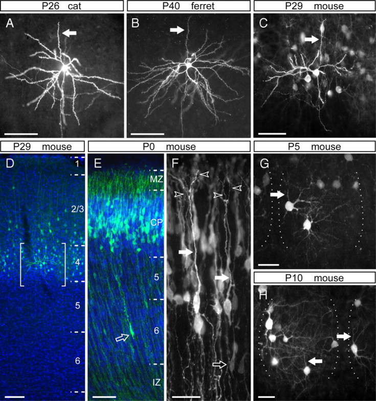
The dendritic arbor of layer 4 cells is dramatically transformed during the first postnatal week in the mouse barrel cortex. A–C, Representative examples of layer 4 spiny stellate neurons in V1 of a P25 cat (A), A17 of a P40 ferret (B), and in the barrel cortex of a P29 mouse (C), in which the apical dendrites are indicated by arrows. D, Cross-section of the barrel cortex of a P29 mouse electroporated at E13.5, in which GFP-transfected cells (green) are positioned in layer 4 and bottom of layer 2/3. Brackets delimit the area shown in C. E, F, Laminar position (E) and detailed morphology (F) of cells in the somatosensory cortex of a newborn mouse, transfected with GFP at E13.5. Bright GFP+ neurons (green in E) align along the deep aspect of the cortical plate (CP) and exhibit a long apical dendrite (filled arrows) with multiple branches within the marginal zone (MZ; open arrowheads). These cells are distinguished from migrating cells in the intermediate zone (IZ) or cortical plate (open arrows) by having a much larger soma (E, F). G, H, Representative layer 4 GFP+ neurons in the barrel cortex of P5 (G) and P10 (H) mice electroporated with GFP at E14.5. These cells display typical stellate morphologies, in which the apical dendrite (arrows) is not more prominent than the basal dendrites. The lateral borders of individual barrels are indicated by dotted lines. DAPI is shown in blue. Scale bars: A–C, E, 50 μm; D, 100 μm; F–H, 25 μm.