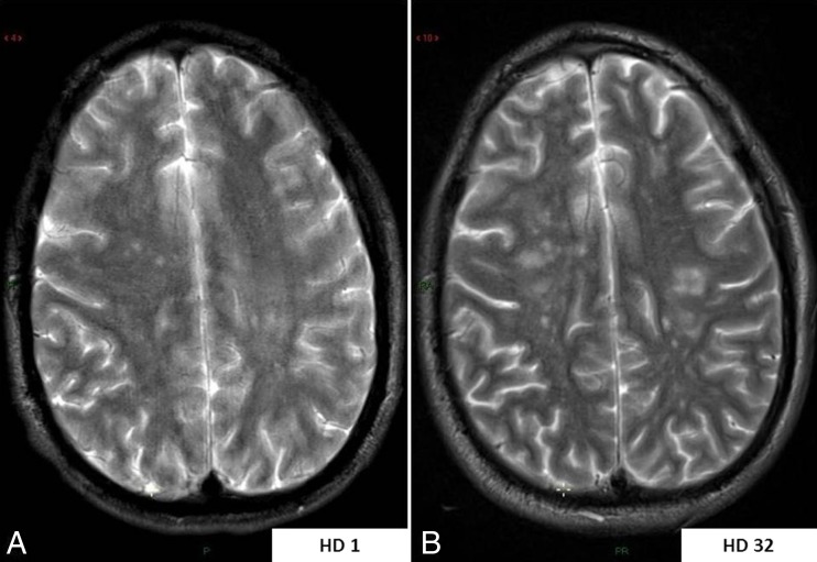
Fig. 3.
MR monitoring of hemispheric white matter lesion load throughout the full disease course. a Axial-transverse T2-weighted image through centrum semi-ovale at initial examination showing only a few aspecific hyperintense white matter lesions within both cerebral hemispheres. b Axial-transverse T2-weighted image in similar slice location at the end of the disease course showing a significant increase in number and size of the white matter lesions

