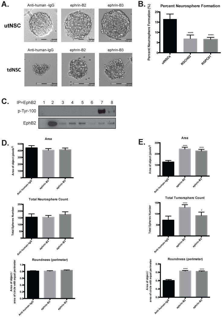
Figure 4. EphB2 driven tumorspheres are uniquely responsive to ephrin-B ligand.
(a) Phase contrast image (20×) of utNSCs (top) and tdNSCs (bottom) after 6-day incubation with anti-human-IgG, ephrin-B2 or ephrin-B3. (b) utNSCs, RGCH02 and RGPC01 sphere formation assay (**** (p < 0.0001) vs. utNSCs). (c) IP/Western of Ink4a/Arf(−/−) STeNSCs (lane 1), utNSCs (lanes 2), tdNSC lines RGPC02, RGCH01-RA, RGPC01-RP, and RGPC03-RM (lanes 3–6), utNSCs treated with ephrin-B2 (lane 7) or anti-human-IgG (lane 8). IP with EphB2 antibody, western blot with phospho-tyrosine (top) or EphB2 (bottom). Graphs of neurosphere (d) and tumorsphere (e) number, area and roundness after treatment with anti-human-IgG, ephrin-B2 or ephrin-B3. * (p < 0.05) and **** (p < 0.0001) vs. anti-human-IgG.

