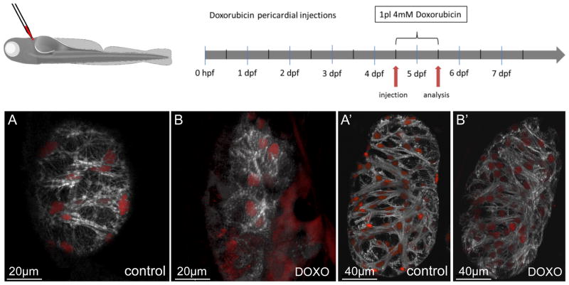
Figure 7. Doxorubicin treatment leads to loss of myofibrils in vivo.
Doxorubicin was injected into the pericardial cavity at 4.5 dpf and the larvae raised for another 24 hours. Subsequent confocal imaging revealed a significant effect on overall myofibril structure and density. While vehicle-injected control larvae showed no detectable alterations (A, A′), injection of 1 pL (4 mmol/L) doxorubicin entirely ablated fine myofibrils in compact wall cardiomyocytes and caused myofibrillar disarray in the thick myofibrils in trabecular cardiomyocytes (B, B′). A′ and B′ are also available as 3-D reconstructions (Online Movies Va and Vb respectively).

