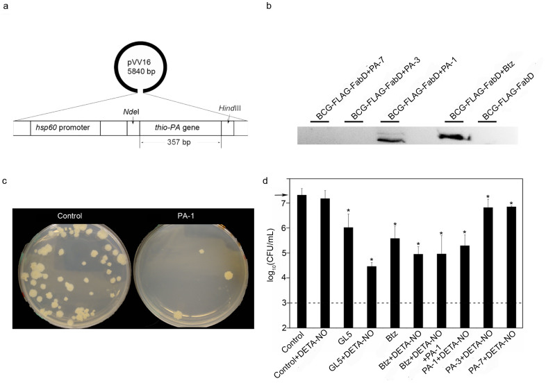
Figure 6. PA-1, but not PA-3 or PA-7, rescues proteasome substrate FabD from degradation and inhibits the growth of BCG.
(a) Diagram of the expression shuttle vector, pVV16, used in the BCG assay. The plasmid contains an E. coli origin derived from pUC19, a mycobacterial origin derived from pAL500046, and a constitutively active BCG hsp60 promoter. (b) BCG-FLAG-FabD was transformed with pVV16-PA-1, -PA-3, and -PA-7 to examine the steady-state level of FLAG-tagged FabD in untreated cells. As a control, BCG was incubated with and without 50 μM Btz. Anti-groEL2 was used as a loading control. (c) BCG plates after 3 weeks of growth show a 100-fold reduction in colonies due to expression of PA-1. (d) Inhibition of BCG growth under conditions that reduce proteasome function. 50 μM Btz, GL5, and DETA- NO were used as indicated. Error bars represent standard deviations and asterisks indicate p- values <0.05 (Prism 5 Graphpad Inc.). The arrow indicates the strength of the initial inoculum. The dashed line is the lower limit of detection. All experiments were performed in triplicate.

