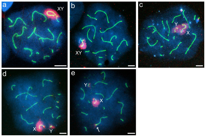
Figure 4. Distribution of γ-H2AFX staining in spermatocyte nuclei.
(a–e) Immunocytochemical staining of γ-H2AFX and the lateral element of the synaptonemal complex in pachytene spermatocytes of Phodopus campbelli (a) and pachytene-like spermatocytes of F1 hybrids (b–e). γ-H2AFX and SYCP3 staining are indicated in red and green, respectively. Spermatocytes with synaptic X and Y chromosomes around which γ-H2AFX was normally distributed (a, b). Hybrid spermatocyte with asynaptic X and Y chromosomes (c–e). The γ-H2AFX distributions were broad (c) or separated (d, e). An autosomal pair was stained with γ-H2AFX antibody (e, arrow). Scale bars represent 10 μm.

